Introduction
The immune system of our bodies has a very tricky mechanism. On one hand, it protects us from external agents, fighting diseases and protecting our healthy cells. On the other hand, though, a slight tweak in the immune system can result in some serious health issues when it starts attacking our own cells. Scleroderma is one such autoimmune disease.
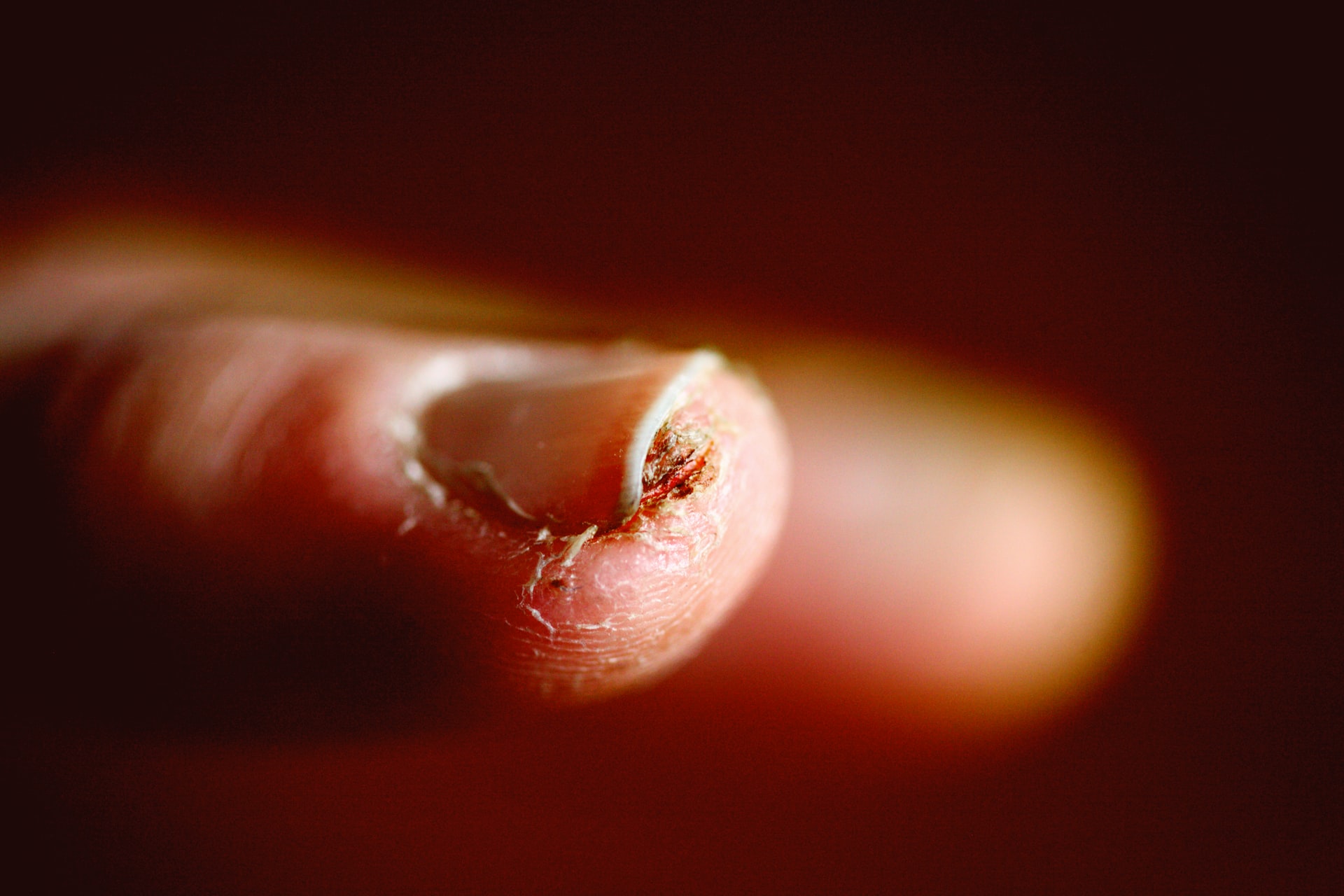
In Scleroderma, our skin, connective tissue, and even our internal organs are affected as the immune system starts producing excessive amounts of collagen. Collagen may be an important substance for the regeneration of the skin, but when it is available in excess, the skin begins to turn thick and hard. These symptoms are mostly seen in the fingers, around the mouth, nose, or other bony areas of the body. It can even lead to more severe symptoms such as scarring of the lungs and kidneys, thickening of the blood vessels, and tissue damage.
Correct diagnosis is the key to managing the disease better and providing the necessary treatment on time. Treatment of the disease works best when started early. But sadly, no medical test is able to confirm that a patient suffers from Scleroderma. Dermatologists have to depend on visible symptoms and maybe a skin biopsy, at best, to determine whether a patient has signs of Scleroderma.
Nailfold Capillaroscopy may have a solution to this problem. Nailfold Capillaroscopy (NFC) is a tool that can make the diagnosis of Scleroderma easier, faster, cheaper, and, most importantly, accurate for physicians. At Capillary.io, we have combined NFC with advanced tools like Machine Learning to help physicians make better decisions in the challenging diagnosis and treatment of Scleroderma.
To understand the crucial role that capillaroscopy can play in the diagnosis of Scleroderma, this article discusses both the disease and the tool in detail.
What Exactly is Scleroderma?
Scleroderma is an autoimmune disorder, in which the immune system attacks the healthy tissue of the body.
There has been extensive research over the years to find out the root cause of the disease but no major breakthrough has been made in this regard. Research has, however, found that women are more commonly affected by the disease than men, but mortality is higher in men. It mainly affects people between 30 and 50 years old, and children are rarely affected. It is also found to affect people of African descent.
A pattern of progression has been found in scleroderma and most frequently the development of Raynaud’s phenomenon (RP) is seen. Raynaud’s phenomenon is a discoloration and numbness of the skin on the fingers and toes. The fingers are first seen to turn white and then worsen to a blue or purple color.
In many cases, digital ulceration and gangrene may also be seen if the disease gets severe.
The types of Scleroderma
There are two types of Scleroderma:
- Localized Scleroderma.
- Systemic sclerosis.
Localized Scleroderma usually affects the skin, muscles, joints, or bone tissues but does not spread to the internal organs. It can further be classified into Morphea and Linear.
In Morphea, red patches appear on the skin that thickens over time to form hard oval-shaped areas. These patches appear on the chest, stomach, back, face, arms, and legs. They may be localized with a few patches, or they may be generalized spread over larger areas, harder and darker in color. Morphea usually fades away in 3 to 5 years, though the dark patches on the skin may remain.
Linear, on the other hand, appears as a thickened band of abnormally colored skin on the arms and legs.
Systemic sclerosis is the more severe form of sclerosis that affects the skin, muscles, joints, blood, vessels, lungs, kidneys, heart, and other organs. The symptoms of systemic sclerosis can be abbreviated as CREST and it is called the CREST syndrome:
- Calcinosis is the formation of calcium deposits in the connective tissue.
- Raynaud’s phenomenon is where the blood vessels of the hands and/or feet contract in response to triggers like cold weather.
- Esophageal dysfunction is the impaired function of the esophageal smooth muscle.
- Sclerodactyly is the thick and tight skin of the fingers due to excess collagen deposition.
- Telangiectasia refers to small red spots on the hands and face due to the swelling of tiny blood vessels.
Systemic sclerosis may be limited or diffuse, each type having slightly different symptoms.
Diagnosis of Scleroderma
Though there is ongoing research, physicians largely depend on certain common symptoms to diagnose Scleroderma, which may include:
- Raynaud’s Phenomenon.
- Skin changes.
- Enlarged red blood vessels on the hands, face, and around the nail beds.
- Calcium deposits in the skin or other areas.
- High blood pressure.
- Heartburn.
- Difficulty swallowing food, bloating, constipation, or issues absorbing food leading to weight loss.
- Shortness of breath.
- Joint pain.
How Nailfold Capillaroscopy can help in the diagnosis of Scleroderma
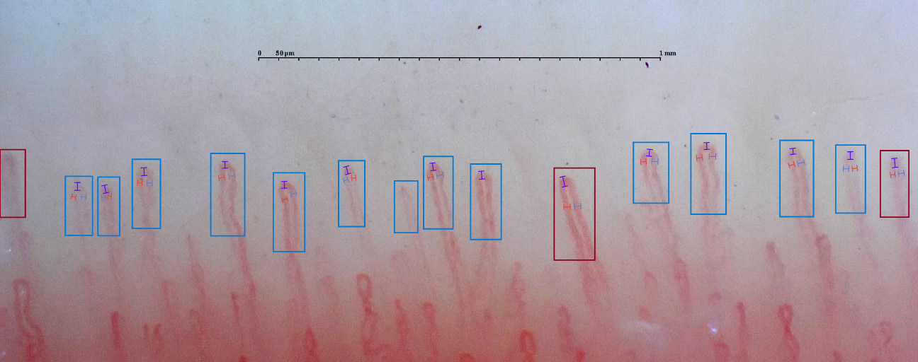
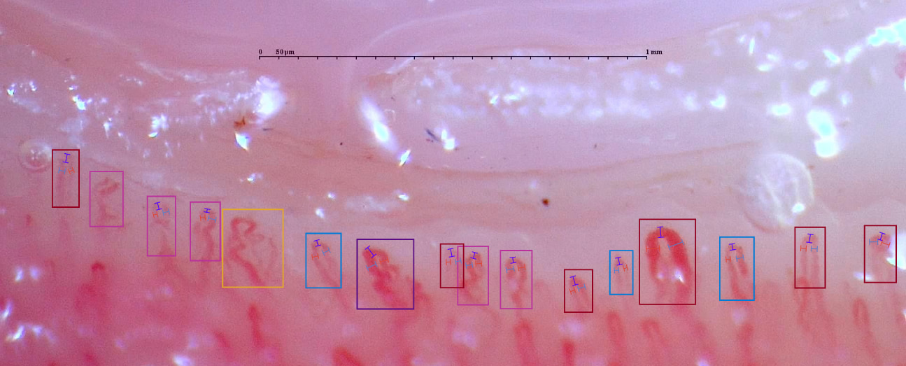
Nailfold Capillaroscopy (NFC) is a non-invasive and inexpensive imaging technique used for diagnosis. In this method, the clinician can view the epidermis of the nailfold to view the nourishing capillaries of the distal papillae.
The biggest advantage of NFC is its ability to detect microvascular changes occurring in inflammatory connective tissue diseases like Scleroderma. This is driving physicians to use NFC as a means of early diagnosis of the disease. It may also be useful in determining which patients have a higher risk of vascular complications and even death from severity in Scleroderma.
Prediction of digital ulcers using NFC
Digital ulcers are a complication in patients suffering from systemic sclerosis. The routine use of NFC in patients has been found to help with the prediction of new digital ulcers. NFC also has uses in identifying pulmonary involvement in patients with lower capillary density.
What else can be detected by Capillaroscopy?
The main indications that can be detected by capillaroscopy include:
- Evaluation of patients with Raynaud’s phenomenon (RP).
- Monitoring the transition from primary to secondary RP.
- Early diagnosis of systemic sclerosis (SSc).
- Differential diagnosis of SSc-related conditions, such as localized SSc and eosinophilic fasciitis.
- Detection of severe microangiopathy and prognostic evaluation in SSc.
- Monitoring of treatment and disease activity in dermatomyositis.
Seeing the importance of NFC in the diagnosis and predictive analysis of Scleroderma, Capillary.io has put in a lot of effort to automate the process, making diagnosis and analysis easier for physicians. The system follows a data-driven approach to NFC by acquiring, storing, and analyzing the metrics for all capillaries on the nailfold. For the specific clinical use case, see Capillary.io for systemic sclerosis capillaroscopy follow-up.
The software can help you decide if there are any changes in the capillaries, using a qualitative and quantitative approach. The collaborative tool also allows sharing capillaroscopy images with other professionals.
Conclusion
The diagnosis of Scleroderma is in need of a revolutionary change. Both physicians and patients suffering from Scleroderma need better and more accurate diagnoses to help start early treatment and prevent severities and life-threatening complications. Nailfold Capillaroscopy has the capability of bringing about this revolution. NFC combined with our cutting-edge solutions could give physicians valuable and deep insights into the disease and aid early detection. Developing the tool has been a challenge for us, but we believe that our platform will transform the way physicians approach scleroderma treatment and improve the standards of care.
Note: If you want to learn about how our tool can help you save time and resources while evaluating patients with scleroderma, you can contact us here.